Preclinical Imaging Center
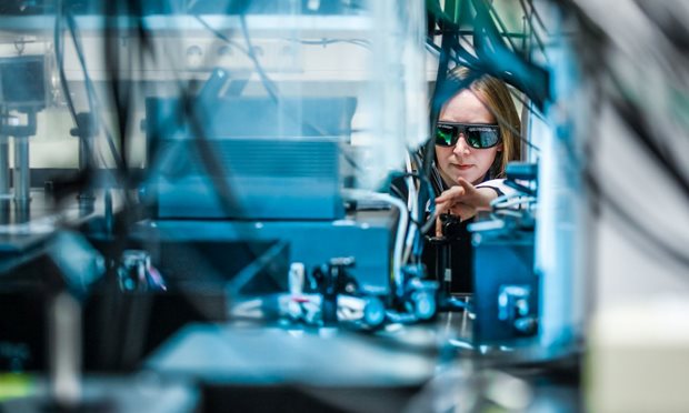
PRIME is a centralized facility for multimodality imaging of small animals, such as mice and rats. Multi modality imaging is a strategic technology, that can be used to support biopharmaceutical development and preclinical research. our aims and servicesPreclinical Imaging Center
Aims
- A durable facility for small animals imaging with state of the art equipment and expertise.
- A facility with multi-modal equipment and offices integrated in one centre at one location.
- A centre that operates essentially as a SPF/DMII unit to allow experiments with transgenic animals and with adeno, retro and lenti-viruses.
- A centre with links to clinical/human imaging facilities.
Services
The infrastructure and imaging equipment in PRIME has some unique features under one roof in one facility:- State-of-the-art scanners for: MRI, SPECT, PET, optical imaging, multiphoton microscopy, intrinsic optical imaging and ultrasound.
- Cognition and behavior rooms to measure (social) behavior learning and memory sensory-motor integration activity and motor skills.
- Barrier-free animal handling at safety level II allowing small animal studies with animals that can be infected with various agents (incl. adeno-/retroviruses, bacteria, parasites).
Contract
PRIME also supports the pharmaceutical and biotechnology industries in the form of research services outsourced on a contract basis. The experts of PRIME can be involved in project development and consultancy as well as giving explanations on the use of our equipment. For more information you can contact Wilma Janssen.PRIME and ...
-
PRIME is a facility that provides support to the pharmaceutical and biotechnology industries in the form of research services outsourced on a contract basis.
read more
Contract Research Organization (CRO)
The Preclinical Imaging Centre (PRIME) is a centralized facility for multimodality imaging of small animals, such as mice and rats. Multi modality imaging is a strategic technology, that can be used to support biopharmaceutical development and preclinical research.
PRIME is staffed by experts from various disciplines such as Radiology, Nuclear Medicine, Cell Biology, Medical Ultrasound Imaging Center, Anatomy and Cognitive Neuroscience. The infrastructure and imaging equipment in PRIME has some unique features under one roof in one facility:- state-of-the-art scanners for: MRI, SPECT, PET, optical imaging, multiphoton microscopy , intrinsic optical imaging and ultrasound.
- cognition and behavior rooms to measure (social) behavior learning and memory sensory-motor integration activity and motor skills.
- barrier-free animal handling at safety level II allowing small animal studies with animals that can be infected with various agents (incl. adeno-/retroviruses, bacteria, parasites).
Investigators from a wide range of research themes use the imaging facilities of PRIME e.g. Alzheimer, cancer development and immune defense, movement disorders, infectious diseases and host response, inflammatory diseases, urological cancers and women's cancers.
PRIME provides research services for third parties outsourced on a contract basis (i.e contract research organization). The experts in PRIME can be involved in project development, consultancy and rental of equipment. For more information you can contact the coordinator of PRIME, Wilma Janssen. -
PRIME is a member of the pan-European Euro BioImaging project which will deploy a distributed biological and biomedical imaging infrastructure in Europa in a coordinated and harmonized manner.
go to Euro BioImaging -
PRIME joined the collaborative DTL platform of life science and technology research groups in the Dutch clinical & health, nutrition, crop and livestock breeding and industrial microbiology sectors.
read more -
PRIME is a member of COMULIS (Correlated Multimodal Imaging in Life Sciences), which is a EU-funded COST Action.
read more
COMULIS
COMULIS (Correlated Multimodal Imaging in Life Sciences) is a EU-funded COST Action that aims at fueling urgently needed collaborations in the field of correlated multimodal imaging (CMI), promoting and disseminating its benefits through showcase pipelines, and paving the way for its technological advancement and implementation as a versatile tool in biological and preclinical research.
For more information, please visit the COMULIS website.
Invitation PRIME lecture
Lecture by Sylvia Wenker and Daniele Taurillo, Monday June 5th at 12:30.
read morePublications in PRIME
1.Lütje S, Franssen GM, Herrmann K, Boerman OC, Rijpkema M, Gotthardt M, Heskamp S. In vitro and in vivo characterization of a 18F-AlF-labeled PSMA ligand for imaging of PSMA-expressing xenografts. J Nucl Med 2019; 60(7):1017-1022.
2.Raavé R, Sandker G, Adumeau P, Borch Jacobsen C, Mangin F, Meyer M, Moreau M, Bernhard C, Da Costa L, Dubois A, Goncalves V, Gustafsson M, Rijpkema M, Boerman O, Chambron J, Heskamp S, Denat F. Direct comparison of the in vitro and in vivo stability of DFO, DFO* and DFOcyclo* for 89Zr-immunoPET. Eur J Nucl Med Mol Imaging 2019; 46(9):1966-1977.
3.Lütje S, Heskamp S, Franssen GM, Frielink C, Kip A, Hekman M, Fracasso G, Colombatti M, Herrmann K, Boerman OC, Gotthardt M, Rijpkema M. Development and characterization of a theranostic multimodal anti-PSMA targeting agent for imaging, surgical guidance, and targeted photodynamic therapy of PSMA-expressing tumors. Theranostics 2019; 9(10):2924-2938.
4.Elekonawo FMK, Lütje S, Franssen GM, Bos DL, Goldenberg DM, Boerman OC, Rijpkema M. A pretargeted multimodal approach for image-guided resection in a xenograft model of colorectal cancer. EJNMMI Res 2019; 9(1):86.
5.Derks YHW, Löwik DWPM, Sedelaar JPM, Gotthardt M, Boerman OC, Rijpkema M, Lütje S, Heskamp S. PSMA-targeting agents for radio- and fluorescence-guided prostate cancer surgery. Theranostics 2019; 9(23):6824-6839.
6.Deken MM, Bos DL, Tummers WSFJ, March TL, van de Velde CJH, Rijpkema M, Vahrmeijer AL. Multimodal image-guided surgery of HER2-positive breast cancer using [111In]In-DTPA-trastuzumab-IRDye800CW in an orthotopic breast tumor model. EJNMMI Res 2019; 9(1):98.
7.Elekonawo FMK, Bos DL, Goldenberg DM, Boerman OC, Rijpkema M. Carcinoembryonic antigen-targeted photodynamic therapy in colorectal cancer models. EJNMMI Res 2019; 9(1):108
8. PSMA-targeting agents for radio- and fluorescence-guided prostate cancer surgery.
Derks YHW, Löwik DWPM, Sedelaar JPM, Gotthardt M, Boerman OC, Rijpkema M, Lütje S, Heskamp S.
Theranostics. 2019 Sep 20;9(23):6824-6839. doi: 10.7150/thno.36739. eCollection 2019. Review.
9. Imaging of T-cells and their responses during anti-cancer immunotherapy.
Krekorian M, Fruhwirth GO, Srinivas M, Figdor CG, Heskamp S, Witney TH, Aarntzen EHJG.
Theranostics. 2019 Oct 16;9(25):7924-7947. doi: 10.7150/thno.37924.
10 .Development and characterization of a theranostic multimodal anti-PSMA targeting agent for imaging, surgical guidance, and targeted photodynamic therapy of PSMA-expressing tumors.
Lütje S, Heskamp S, Franssen GM, Frielink C, Kip A, Hekman M, Fracasso G, Colombatti M, Herrmann K, Boerman OC, Gotthardt M, Rijpkema M.
Theranostics. 2019 May 4;9(10):2924-2938. doi: 10.7150/thno.35274. eCollection 2019.
11. CAIX-targeting radiotracers for hypoxia imaging in head and neck cancer models.
Huizing FJ, Garousi J, Lok J, Franssen G, Hoeben BAW, Frejd FY, Boerman OC, Bussink J, Tolmachev V, Heskamp S.
Sci Rep. 2019 Dec 11;9(1):18898. doi: 10.1038/s41598-019-54824-5.
12. Tengeler AC, Dam SA, Wiesmann M, Naaijen J, van Bodegom M, Belzer C, Dederen PJ, Verweij V, Franke B, Kozicz T, Arias Vasquez A, Kiliaan AJ. Gut microbiota from persons with attention-deficit/hyperactivity disorder affects the brain in mice. Microbiome. 2020 Apr 1;8(1):44. doi: 10.1186/s40168-020-00816-x. PubMed
PMID: 32238191; PubMed Central PMCID: PMC7114819.
13. Calahorra J, Shenk J, Wielenga VH, Verweij V, Geenen B, Dederen PJ, Peinado MÁ, Siles E, Wiesmann M, Kiliaan AJ. Hydroxytyrosol, the Major Phenolic Compound of Olive Oil, as an Acute Therapeutic Strategy after Ischemic Stroke. Nutrients. 2019 Oct 11;11(10). pii: E2430. doi: 10.3390/nu11102430. PubMed PMID: 31614692;
PubMed Central PMCID: PMC6836045.
14.Jacobs SAH, Gart E, Vreeken D, Franx BAA, Wekking L, Verweij VGM, Worms N, Schoemaker MH, Gross G, Morrison MC, Kleemann R, Arnoldussen IAC, Kiliaan AJ. Sex-Specific Differences in Fat Storage, Development of Non-Alcoholic Fatty Liver Disease and Brain Structure in Juvenile HFD-Induced Obese Ldlr-/-.Leiden Mice. Nutrients. 2019 Aug 10;11(8). pii: E1861. doi: 10.3390/nu11081861. PubMed PMID:
31405127; PubMed Central PMCID: PMC6723313.
15. van Heukelum S, Mogavero F, van de Wal MAE, Geers FE, França ASC, Buitelaar JK, Beckmann CF, Glennon JC, Havenith MN. Gradient of Parvalbumin- and Somatostatin-Expressing Interneurons Across Cingulate Cortex Is Differentially Linked to Aggression and Sociability in BALB/cJ Mice. Front Psychiatry. 2019 Nov 15;10:809.
16. Jager A, Kanters D, Geers F, Buitelaar JK, Kozicz T, Glennon JC. Methylphenidate Dose-Dependently Affects Aggression and Improves Fear Extinction and Anxiety in BALB/cJ Mice. Front Psychiatry. 2019 Oct 25;10:768.
17. Havenith MN, Zijderveld PM, van Heukelum S, Abghari S, Tiesinga P, Glennon JC. The Virtual-Environment-Foraging Task enables rapid training and single-trial metrics of rule acquisition and reversal in head-fixed mice. Sci Rep. 2019 Mar 18;9(1):4790.
18 . Odenthal J, Friedl P, Takes RP. Compatibility of CO2 laser surgery and fluorescence detection in head and neck cancer cells. Head Neck. 2019 May;41(5):1253-1259.
19. Weigelin B, den Boer AT, Wagena E, Broen K, Dolstra H, de Boer RJ, Figdor CG, Textor J, Friedl P. Cytotoxic T cells are able to efficiently eliminate cancer cells by additive cytotoxicity. Nat Commun. 2021 Sep 1;12(1):5217.
New equipment in PRIME
At PRIME, a high-power portable fiber coupled diode laser is available. read moreNew equipment in PRIME
In targeted photodynamic therapy, molecules named photosensitizers are brought to tissues or cells of interest. These photosensitizers can be activated with light of a specific wavelength, upon which they generate the toxic reactive oxygen species and induce cell death.
At PRIME, a high-power portable fiber coupled diode laser is available (BWF2 B&WTek inc). It emits 690 nm light with an adjustable output power up to 500 mW. Furthermore a collimating lens is available, which makes the system suitable for homogenous illumination of a 5 mm diameter surface. Using this system, NIR-photosensitizers such as the frequently used IRDye700DX can effectively be activated.
MRI scanner for mice
Less need for animal testing thanks to MRI scanner for mice. read more on NOSImaging technologies
Powerful techniques in biomedical research
All pre-clinical non-invasive imaging technologies available in PRIME are powerful techniques in biomedical research. Combining the strengths of anatomical and functional imaging modalities allows the detection and quantitative determination of morphological, physiological and metabolic processes.
Facility MR imaging
An MR system uses a strong magnetic field to align the magnetization of some atoms in the body, and radio frequency fields to systematically alter this alignment for detection purposes. read more
Facility Radionuclide imaging
Radionuclide imaging relies on the tracer principle in which a minute amount of a radioactive tracer is injected into the body to monitor a physiological/biochemical process. read more
Facility Optical imaging
Optical imaging offers unique possibilities for in vitro and in vivo imaging applications, especially in the context of molecular imaging. read more
Facility Ultrasound and photoacoustic imaging
High Frequency Ultrasound and Photoacoustic Imaging is a technique used to visualize detailed internal structures and function with unprecedented temporal and spatial resolution. read more
Facility Multi-photon microscopy
Multi-photon excitation microscopy (MPM) is the approach of choice for fluorescence imaging of living three-dimensional tissue at subcellular resolution for up to one millimeter in depth. read more
Facility Behavior and cognition
In the Behavior & Cognition Facilities, (social) behavior, cognition, sensory-motor integration and blood pressure in neural development, adolescence, ageing and neurodegenerative diseases can be measured in several mice models. read more
Facility Intrinsic optical and multi-photon imaging
The combination of intrinsic optical imaging and multiphoton microscopy has many applications in neuroscience, because it allows to measure neural activity and structural changes in the nervous system at multiple scales. read more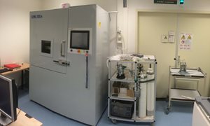
Facility Radiotherapy
With our XRad we can irradiate tumors in animals with varying doses. Our Physicists and technicians will determine the settings of the equipment and after that treatment with a dose varying from almost zero to >50Gy is possible. read more-
Detecting and treating tumor cells during prostate cancer surgeryDefense of Yvonne Derks' thesis on 8 September8 September 2022
-
Liver tumors irradiated from inside can be followed live from outside17 February 2022
-
LUMO Labs and Oost NL invest in AiosynInvestment accelerates development of artificial intelligence platform to improve diagnostics7 February 2022
Getting there
Visiting address
PRIME
Geert Grooteplein 29
6525 EZ Nijmegen
Directions


-
Alex Hanssen senior biotechnicus
-
Wilma Janssen-Kessels procescoördinator PRIME
-
Floor Moonen biotechnicus
-
Liz van den Brand teamcoördinator CDL
Departments
PRIME is a partnership between the departments that provide dedicated state-of-the-art equipment and expertise.- Radiology
- Nuclear Medicine
- Cell Biology
- Anatomy
- Cognitive Neuroscience

Radboudumc Technology Center Imaging
This technology center provides cutting-edge technology and service for imaging-related preclinical and clinical research questions. This covers the spectrum from molecule to man to population.
read more
Radboudumc Technology Center Animal research facility
This technology center offers advice and support from planning up to and including the conduct of animal research on behalf of biomedical research and education.
read more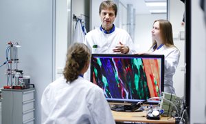
Radboudumc Technology Center Microscopy
This technology center offers access to both standard and innovative advanced microscopy infrastructure and applications including technical operator-assistance and in-depth microscopic imaging knowledge.
read more


