About our equipment
Multi-photon excitation microscopy (MPM) is the approach of choice for fluorescence imaging of living three-dimensional tissue at subcellular resolution for up to one millimeter in depth. read moreAbout our equipment
Multi-photon excitation microscopy (MPM) is the approach of choice for fluorescence imaging of living three-dimensional tissue at subcellular resolution for up to one millimeter in depth. The PRIME Nijmegen MPM setup consists of two TriMScope-II microscopes (LaVision Biotec, Germany), it incorporates three fs-pulsed excitation sources (Chameleon Ultra, Coherent) and an optical parametric oscillator (Coherent-APE) to excite samples with two near infrared and one infrared wavelength simultaneously.Technical Details about MPM
The MPM in PRIME has up to 7 non-descanned detection channels available in the backward and forward direction. read moreTechnical Details about MPM
The MPM in PRIME has up to 7 non-descanned detection channels available in the backward and forward direction, including two 16-channel time domain fluorescent lifetime detectors for intravital fluorescent lifetime imaging. Infrared (IR-)MPM reaches up to 10-fold improved excitation of far-red fluorophores (e.g. DsRed, mCherry), two-fold tissue penetration, reduces photobleaching 4-10 fold and minimizes phototoxicity, compared with conventionally used near-infrared excitation (Andresen et al. , 2009), thus maximizing sample volume and biocompatibility during intravital microscopy.
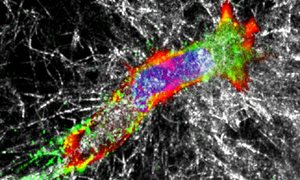
Visualization of signals with MPM
IR-MPM in PRIME allows to visualize several types of signals from live tissues by simultaneous detection. read moreVisualization of signals with MPM
IR-MPM in PRIME allows to visualize several types of signals from live tissues by simultaneous detection:- Fluorescence intensity of blue, green, red and far-red probes and proteins;
- Second harmonic generation by collagen and actomyosin in mascle fibers;
- Third harmonic generation of interfaces between (bio)materials with different optical properties, including nerve cells, blood cells and intra-extracellular boundaries (e.g. lipid bodies, cell fragments);
- Fluorescence lifetime of fluorescence emission from single- and multi-spectral samples, including autofluorescent drugs and molecular FRET/FLIM reporters.
