About our equipment
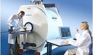
An MR system uses a strong magnetic field to align the magnetization of some atoms in the body, and radio frequency fields to systematically alter this alignment for detection purposes. read moreAbout our equipment
An MR system uses a strong magnetic field to align the magnetization of some atoms in the body, and radio frequency fields to systematically alter this alignment for detection purposes. Using the signals of hydrogen in water, MRI routinely provides good contrast between and within soft tissues in the body. This contrast can be varied for optimal visualization (e.g. of diseased tissue).On top of this MR can be used for bone and (large) vessel imaging and to obtain functional tissue information such as vascular parameters (blood volume, flow), metabolism, oxygenation, cell density and more. Unlike CT or traditional X-rays, MRI uses no ionizing radiation, but stable isotopes or contrast media can be used to track certain processes or visualize tissues and targets.
In the PRIME facility two MR systems with horizontal bore magnets are available: one at 7T field strength and another at 11.7T. Both systems have a large range of radiofrequency probes, mostly focusing on mice and rat applications (e.g. for muscle, brain). Both systems have anesthesia and animal monitoring devices.
Applications in PRIME
The 7 and 11.7 T scanners in PRIME can be used in several applications. All applications can performed quantitative. read moreApplications in PRIME
The 7 and 11.7 T scanners in PRIME can be used in the following applications. All applications can performed quantitative.- Anatomy and morphology;
- Metabolism, i.e. metabolite mapping with spectroscopic imaging;
- Metabolism with endogenous isotypes or stable isotope labels (1H, 31P, 13C, etc.);
- Perfusion and vascular imaging;
- Diffusion and diffusion tensor imaging;
- Cellular imaging;
- Targeted contrast MR, e.g. in cellular imaging;
- Quantitative blood flow.


