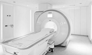
Why an MRI-scan?
An MRI scan uses a strong magnetic field and radio waves (not X-rays) to create pictures, on a computer, of tissues, organs and other structures inside your body. An MRI scan uses a strong magnetic field and radio waves (not X-rays) to create pictures, on a computer, of tissues, organs and other structures inside your body.MRI Checklist
Please read and complete the following checklist before your appointment. read moreThe examination
Before the examination
Before the examination, some precautions should be taken. Moreover, some preparations should be taken care of.
read more
During the examination
You are required to report to the Radiology Department (Route 780) 10 minutes before the time stated on your appointment card or letter, keeping in mind that it’s a 10 minute walk from the main entrance to the Radiology Department. read moreDuring the examination
You are required to report to the Radiology Department (Route 780) 10 minutes before the time stated on your appointment card or letter, keeping in mind that it’s a 10 minute walk from the main entrance to the Radiology Department. Should you require assistance with mobility, transportation can be arranged at the reception
desk in the main hall.
The radiographer or assistant will collect you (and your companion) from the waiting area, bring you to a changing room where you (and your companion) will be required to leave all metal objects, telephones, credit-bank cards etc, behind. If necessary an intravenous line will be inserted and the procedure will be explained.
You may direct any questions to the radiographer at any time before or during the preparation for your examination.
When you enter the MRI examination room, you will be required to lie on the scanner table. To detect the tiny radio signals that are emitted from the body, a ´receiving device´ is placed behind or around the area to be examined. You will be given a rubber ball to hold onto during the scan. This is the alarm bell and the radiographer will instruct you as to when you can use it.
MRI has been shown to be extremely safe as long as proper safety precautions are taken. In general, the MRI procedure produces no pain and causes no known short-term or long-term tissue damage of any kind.
The part of your body being examined is placed in the middle of the scanner (an open-ended, cylinder-shaped machine about a metre long). During the examination you will hear different kinds of loud noises (knocking/buzzing) but will be given earplugs or headphones to wear. If you wish you may listen to the radio or a cd. During the examination many images are taken. Some images only take a few seconds to make, others several minutes. Once the noise has stopped, then one set of images have been made. During the making of the images and the time in between, you will be required to lay very still. Once all the images have been made the radiographer will slide you out of the scanner, and if you had a contrast agent, will remove the intravenous line. The whole examination takes on average, around 30 minutes, but in some cases can take up to an hour or longer.
Contrast Agents
It is possible that a contrast agent may be administered during your scan. This is determined by the specialist and radiologist and allows the differences between organs and tissues on an MRI scan to be seen more clearly. To administer the contrast agent the radiographer will insert an cannula/intravenous line and will remove the cannula at the end of the examination. During the examination the radiographer will administer the contrast agent. Usually, this will pose no discomfort however you may experience a cold sensation in the arm or a strange taste in the mouth. These sensations only last a few seconds. Rarely do patients have a reaction to the contrast agents used for MRI scans, but should you have had a reaction in the past, please contact the Radiology Department as the radiologist can in consultation with your specialist, take preventative measures. If you have a reduced kidney function we would also request that you contact the Radiology Department.
