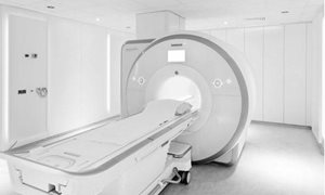
About the MRI-guided biopsy of the prostate
You have already had an MRI scan (Magnetic Resonance Imaging) of your prostate gland whereby some irregularities have shown up. To be able to determine what exactly these irregularities are, your specialist (Urologist/Radiotherapist) has requested a second MRI examination. read moreAbout the MRI-guided biopsy of the prostate
You have already had an MRI scan (Magnetic Resonance Imaging) of your prostate gland whereby some irregularities have shown up. To be able to determine what exactly these irregularities are, your specialist (Urologist/Radiotherapist) has requested a second MRI examination. During this second examination, visible
abnormalities can be located and small samples can be taken for further analysis by a pathologist (biopsy). This folder will explain the procedure.
On this website, how the examination is carried out is only a general description of a procedure. It may be that your specialist requests a procedure that may vary from the one described here, as it is not possible to list every variant of the procedures that are carried out. Risks and side-effects are only explained in general terms and possible complications will be explained by your specialist.
Your appointment
Your doctor will make an appointment. You will get a notification. If you are unable to attend, please let us know as soon as possible so that your appointment can be given to another patient. read moreYour appointment
Your doctor will make an appointment. You will get a notification.Cancelling or changing your appointment
If you are unable to attend, please let us know as soon as possible so that your appointment can be given to another patient. You can call the Radiology Department on weekdays between 08.30-16.45 at (024) 361 45 29.Should you wish to cancel your appointment we request that you also inform your specialist.
Preparation
Unless otherwise requested by your specialist, you may continue to take any medications and eat and drink normally. Rings and piercings made of gold or silver may be worn as the magnet doesn’t affect these items. read more
During the procedure
You will be required to lie on the scanner table, head first and on your front (see illustration). The attending physician will first feel for the position of the prostate gland through the anus. read moreDuring the procedure
You will be required to lie on the scanner table, head first and on your front (see illustration). The attending physician will first feel for the position of the prostate gland through the anus. They will then insert a thin tube (called a needle guide) into position in the rectum which is covered with a lubricant containing pain-relieving properties, to make the insertion easier. The tube will then be connected to the biopsy apparatus which is used to adjust the position of the needle guide. Antenna in the form of a plate (also used in the previous MRI scans) will be placed over your back.A number of scans are then made to produce images of your prostate and to give the exact location that the needle will be inserted into. The table will be slid out of the scanner several times while the position of the needle guide is adjusted so that it is directed to the region where the samples are to be taken. Once the physician is satisfied that the needle guide is in the correct position, the biopsy needle is inserted and the biopsy will be performed. The taking of these samples may be uncomfortable. A control scan is made, to check the position of the needle.
Generally, 2 to 3 samples are taken from each suspicious area. It may be necessary to take further samples from other areas in the prostate gland.
The whole procedure takes between 40-50 minutes.
