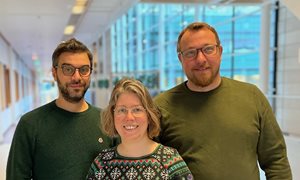
Ioannis Sechopoulos has been appointed professor of Advanced X-ray Imaging Methods at Radboud university medical center / Radboud University. Sechopoulos works on improving imaging with x rays, particularly for breast cancer applications. He improves both the equipment and the analysis of x-ray images. He is also investigating in clinical trials the value of improved imaging methods in cancer screening and treatment, and for other diseases.
Two years ago, Ioannis Sechopoulos started a project to create a new x-ray imaging method; 4-dimensional breast CT. This new technique will be used on breast cancer patients. With this technique, doctors will better understand the breast tumor and improve its treatment. Next year, once the CT system is ready, Sechopoulos will perform two clinical trials to evaluate its use. It's a great example of Sechopoulos' work: first creating or improving a technique and then testing what it brings to patients.
Sechopoulos was already appointed professor at the University of Twente in 2021. There, he tinkers with the x-ray equipment itself. For example, with new X-ray sources or improved detectors that capture and measure the X-rays. At the Radboudumc, Sechopoulos is working on follow-up steps, such as improving the creation and analysis of images with the help of computers. He is also conducting clinical studies.
Patterns
According to Sechopoulos, we can extract much more information from x-ray images: 'We now mainly look at the shape and size of an abnormality, for example in the breast. If we want to know more about the type of tissue that makes up the abnormality, we usually opt for an MRI or PET scan. But those methods are expensive and slow. Whereas you can also get that information with smarter creation and analysis of x-ray images.’
This is how it works: Sechopoulos makes many images, or even a kind of 4-dimensional movie, with x rays. This movie shows how the blood flows in the tumor. Using AI, he will look for patterns in the blood flow. Based on that, the AI may provide more information about the type of tumor tissue, even down to genetic and molecular characteristics. That information could help choose the right treatment.
Structure
Such analysis with CT could potentially even prevent surgery in the future. ‘One biopsy is needed to confirm cancer with pathology’, says Sechopoulos. 'But tumors are always heterogeneous, meaning that the tissue in the tumor is not the same everywhere. With advanced x-ray imaging, you can assess the rest of the tumor without taking more biopsies. And you can also follow a tumor during therapy: if you see a tumor disappear, surgery may no longer be necessary.'
For conditions other than breast cancer, it is also interesting to use CT to look at the composition and behavior of tissues, in addition to size and shape. For example, Sechopoulos also works on lung cancer and heart disease.
Career
Ioannis Sechopoulos (Athens, 1975) received his MSc in Mechanical Engineering from Stanford University. He received his PhD from the Georgia Institute of Technology, on his dissertation entitled: Investigation of Physical Processes in Digital X-ray Tomosynthesis Imaging of the Breast. Sechopoulos worked at Emory University School of Medicine and at University of Massachusetts Medical Center. Since 2015, he has worked at Radboudumc.
Sechopoulos received many grants, including NIH RO1s, ERC consolidator, KWF and NWO Talent (VICI) and has been named Fellow of the American Association of Physicists in Medicine. In 2021, Sechopoulos was already appointed professor at the University of Twente. The appointment as professor at Radboudumc will take effect July 1, 2023 for a period of five years.
-
Want to know more about these subjects? Click on the buttons below for more news.
More information
Annemarie Eek

wetenschapsvoorlichter


