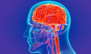
Researchers from Radboud university medical center have developed a set of growth charts for the brain. These ‘brain charts’ provide reference models for brain development and ageing across the entire human lifespan, based on a very large data set. These models can be used to make personalized predictions for each individual relevant to many brain conditions, and therefore have a high clinical potential. The software tools and models are available online. The work has been published in eLife.
“Nearly everybody is familiar with the growth charts used to measure child development, for example the growth charts developed by the World Health Organization,” says Andre Marquand, a researcher at the department of Cognitive Neuroscience of Radboud university medical center. “These models are being used worldwide to assess the development of children, for instance by plotting body weight or height as a function of age. Pediatricians plot the development of an individual child against variation in the population provided by these growth charts, in order to detect, for example, developmental delay.”
The researchers now provide the same thing for the brain: a growth chart to assess brain development and aging, not only for children, but across the lifespan from ages two to 100. “We have analyzed high resolution MRI images from nearly 60.000 people from around 80 MRI scanners all over the world,” explains Saige Rutherford, PhD candidate and first author. “We used measures of the volume of different brain structures or the thickness of the cerebral cortex at different ages and created growth charts for every brain region. In this way, we created a fine-grained atlas of the human brain throughout life.”
Alterations in brain structure
These models enable predictions at the level of an individual person about brain growth and ageing, with respect to population norms. Marquand: “This provides a reference model to map variation across individuals and can be used to help understand many different brain-based conditions, like ADHD, schizophrenia, dementia and Alzheimer’s disease.”
These models have many uses: they can be helpful to detect alterations in brain structure that might indicate the emergence of a mental disorder at a very early stage. The models can assess if a region in the brain is thicker or thinner than it ought to be for an individual compared to the average for this life stage. But it is also useful for stratification of mental disorders. For example, finding commonalities between individuals that might describe different subtypes of disorders, or in the future to identify individuals that could respond to certain treatments. In addition, the model enables tracking of disease progression over time, and also monitoring the effect of a treatment.
Brain fingerprints
A reference model for the brain like this has not been available before. The models and also the software to use them are made freely available online to the community. “We use an established software pipeline called ‘Freesurfer’ to measure the volume and thickness of brain structures,” explains Marquand. “This pipeline is used by thousands of hospitals worldwide, so they can easily get the measures they need and use our software to determine how a group of their own patients or study participants can be placed within the population.”
In the near future, Marquand thinks the software could be of great use in clinical studies. “If you want to investigate a new medication against a certain brain-based condition, for example Alzheimer’s disease, you could use our software to identify subjects with a particular profile, such as early stage degeneration. This could function like a ‘brain-based fingerprint’ which could make research more efficient by making it easier to detect differences between groups of people. Eventually, such tools might also be helpful in the clinic to target medications or interventions precisely to the people that need them.”

Example of a growth chart developed by the researchers. This growth chart shows the volume of the amygdala, a region of the brain frequently associated with emotions. The amygdala was measured with MRI in many individuals at different ages. All data were plotted and show the average volume of the amygdala and the variation in the population from ages two to 100.
About the publication
This research was published in eLife (link): Charting Brain Growth and Aging at High Spatial Precision. Saige Rutherford, Charlotte Fraza, Richard Dinga, Seyed Mostafa Kia, Thomas Wolfers, Mariam Zabihi, Pierre Berthet, Amanda Worker, Serena Verdi, Derek Andrews, Laura Han, Johanna Bayer, Paola Dazzan, Phillip McGuire, Roel T. Mocking, Aaart Schene, Brenda W. Pennix, Chandra Sripada, Ivy F. Tso, Elizabeth R. Duval, Soo-Eun Chang, Mary Heitzeg, S. Alexandra Burt, Luke Hyde, David Amaral, Christine Wu Nordahl, Ole A. Andreasssen, Lars T. Westlye, Roland Zahn, Henricus G. Ruhe, Christian Beckmann, Andre F. Marquand.
-
Want to know more about these subjects? Click on the buttons below for more news.
More information
Annemarie Eek

wetenschapsvoorlichter
Related news items

Dogma broken: sex differences in XLMTM mapped out Women also experience muscle symptoms due to genetic disorder X-linked MTM
6 October 2022For a long time, healthcare professionals thought that only men could suffer from XLMTM, a serious muscle disease that is inherited via the X chromosome. It now appears that women with this genetic defect are also prone to this disease.
read more


