Radboud university medical center and Radboud University, together with the universities of Leiden and Amsterdam, will use electron microscopy to obtain new insights and leads for the development of better medicines. To establish the required research facilities, Nico Sommerdijk and colleagues will both receive an NWO-Groot grant of two 1.5 million euros and an additional contribution of 500 thousand euros from Radboudumc.
The complex biological processes inside and outside our cells form the basis of both health and disease. The development of new therapies for major diseases such as cancer, Alzheimer's and osteoporosis requires a fundamental understanding of these processes. In search of new fundamental insights and leads for the development of better medicines, researchers Nico Sommerdijk and Niels de Jonge, at the forefront of this field, are introducing a new type of electron microscope to study biological systems real time - without damaging them - in their natural habitat in detail. This nanoscopic imaging of biological systems sensitive to electron beams is challenging, but essential for improved understanding in the life sciences.
Nobel Prizes for Microscopy
Recent innovations in biological microscopy techniques have already advanced the field, as highlighted by the two Nobel Prizes awarded for the development of cryo-electron microscopy and super-resolution microscopy. Yet, these techniques are not able to reveal the full dynamics and context of the monitored processes, or these techniques lack the required spatial resolution. Recent developments in liquid phase electron microscopy have opened up the possibility to finally overcome these limitations and provide a powerful tool for dynamic imaging of biological processes the molecular scale.
National facility to better understand biological systems
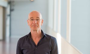
Nico Sommerdijk, Professor of Bone Biochemistry at Radboud university medical center, explains his research. “We are going to set up a globally unique facility using the recently awarded NWO Groot (BIOMATEM). This facility will be the first of its kind dedicated to dynamic electron microscopy of biological materials in liquid, and as such will be internationally attractive for many researchers to conduct pioneering research. The technology is already highly advanced and can be applied immediately to achieve breakthroughs in the research programs of several Dutch biomedical groups. At the same time, ongoing innovations in hardware, software and sample preparation will significantly improve the imaging quality, expanding the variety of biological systems that can be examined.”
About the BIOMATEM project
BIOMATEM stands for In Situ Imaging of Biological Materials with Nanoscale Resolution using Liquid Phase Electron Microscopy. Co-applicants from Nijmegen are Dr. Elena Macias, Prof. Niels de Jonge, Prof. Sander Leeuwenburgh and Prof. Daniela Wilson (Radboud University).
About NWO Groot
With this program the Dutch scientific organization NWO funds large scientific facilities in which scientists from across the Netherlands collaborate, often with international partners. This investment enables a wide range of researchers to conduct innovative experiments and unlock new data. By making these investment funds available for the purchase of high-quality equipment, data collections and software, NWO strengthens the scientific infrastructure of Dutch knowledge institutions. With these funds NWO supports large national and international scientific projects, thereby also stimulating cooperation between scientists.
-
Want to know more about these subjects? Click on the buttons below for more news.
More information
Pauline Dekhuijzen

wetenschaps- en persvoorlichter
Related news items

Rubicon grants awarded to three RIMLS researchers
19 April 2022Three researchers have received Rubicon funding from NWO/ZonMw. This will enable Elke Muntjewerff, Laura de Vries and Laurens van de Wiel to do research at a foreign research institute for the next two years.
read more
Large NWA ORC grant awarded for national skin research: Next Generation ImmunoDermatology
23 March 2022Research for better treatment methods for chronic skin diseases.
read more


